
Other important information to discuss with the doctor before the procedure includes the patient’s medical history and adverse reactions to prior medical imaging procedures. They must also disclose if they are claustrophobic and in need of sedation. Patients must inform their radiologist if they have any allergies to contrast dyes. What to Expect From a Thigh MRI Before the ProcedureĪ patient undergoing thigh MRI may follow their routine and take medication as usual unless their physician advises otherwise. An MRA may also help surgeons prepare for surgery on the arteries of the legs(9). Other types of MRI, such as magnetic resonance angiogram (MRA), may capture medical images of the body’s blood vessels and blood flow(8).ĭoing an MRA of the legs may help physicians detect stenosis (narrowing) and blockage of the arteries, also known as peripheral arterial disease. It involves magnetic fields and radio waves to develop images of the body’s internal organs(7). Magnetic resonance imaging (MRI) is a medical imaging procedure that may be used to diagnose conditions of the legs. The rectus femoris is also able to flex the thigh at the hip(6). The four muscles all extend the lower leg. Meanwhile, the vastus lateralis is on the side of the thigh, while the vastus intermedius is hidden below the rectus femoris(5). The rectus femoris is located in the center of the thigh, while the vastus medialis is in the middle of the said body part. The thigh is composed of several muscles, including the quadriceps or quads (a group of four muscles)(4): Thigh muscles also protect neurovascular structures as they go through the proximal hip joint to the knee and lower leg(3). Weak adductor muscles may cause knee instability and adductor strain(2). The medial thigh muscles are responsible for the adduction (movement of a body part toward the body’s midline) of the leg.

Thigh muscles are responsible for allowing normal gait and proper lower extremity function(1).

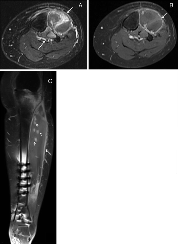
The thigh has some of the body’s largest muscles. In view of the substantial increase in T2-weighted signal intensity, MRI can be used in diagnosing chronic compartment syndrome.This webpage presents the anatomical structures found on thigh MRI. This effect disappeared after fasciotomy. In patients with a chronic compartment syndrome, the affected (anterior) compartment shows a statistically significant increase in (T2-weighted) signal intensity during exercise compared with both the (superficial) posterior compartment and the anterior compartment of normal controls. Following fasciotomy, the increase in the anterior compartment was 4.1% (range 1.0–5.2%), while the increase in the posterior compartment amounted to 5.6% (range 0–11.0%), In normal controls, the increase in the anterior compartment was 7.6% (range 0–9.1%), while in the posterior compartment it was 4.0% (range 0–7.2%).Ĭonclusions. In the posterior compartment this increase amounted to 4.25% (range 0–10.2%). T 2-weighted signal intensity increased by 27.5% (range 13.6–38.6%) following exercise in the anterior compartment of patients with a chronic compartment syndrome. MR studies were performed in 12 normal controls (24 anterior muscle compartments) on the basis of the same protocol. After fasciotomy, a second MRI scan was performed in 13 patients (25 anterior compartments) on the basis of the same protocol. Postexercise increases in the signal intensity in these two compartments were compared. Median (T2-weighted) signal intensity on the MRI scan was determined in the anterior and the (superficial) posterior compartment of the lower leg before and after exercise. MRI was performed in 21 patients (41 anterior compartments) with chronic compartment syndrome at rest and following physical exercise. A prospective descriptive study to determine the value of magnetic resonance imaging (MRI) as an aid in diagnosing (chronic) exertional compartment syndrome.ĭesign and patients.


 0 kommentar(er)
